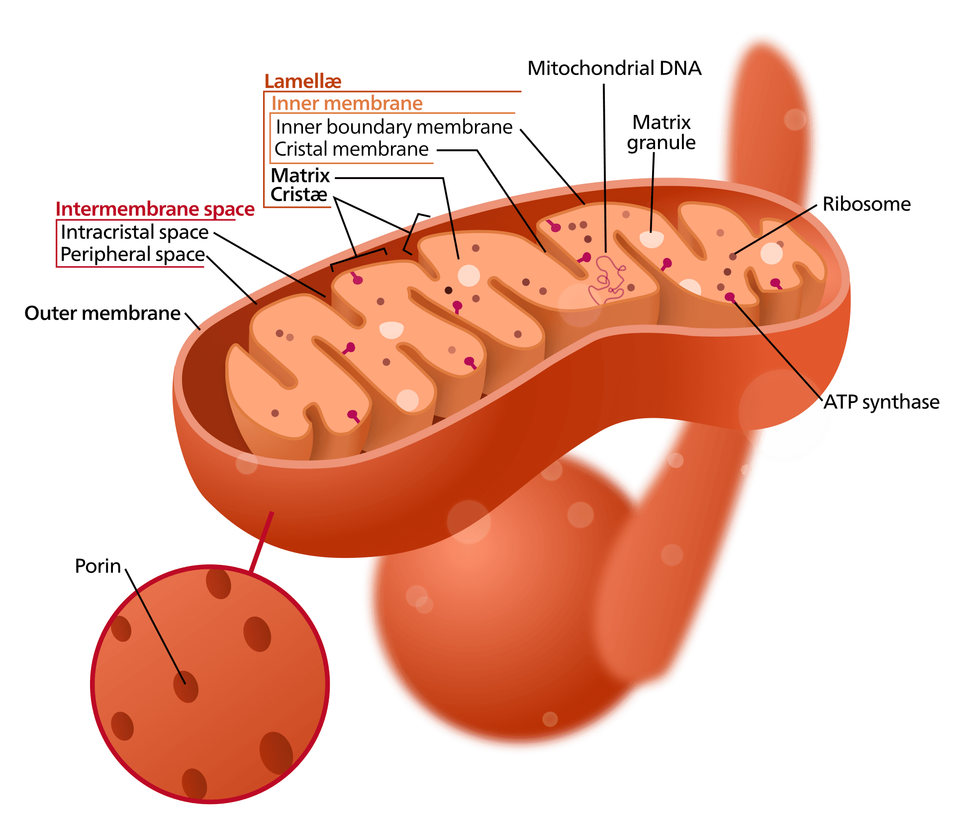
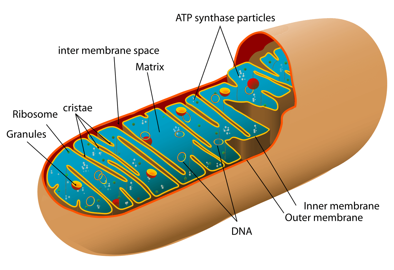
Inside each human muscle cell, a microscopic marvel unfolds. Approximately 1,500 mitochondria are neatly arranged along the cell's fibres, each a mere micrometre in size. Far from being mere cellular components, these mitochondria possess their own membranes, ribosomes distinct from those of their host cell, and a genome of 37 genes configured in a circular chromosome—reminiscent of bacterial DNA rather than the linear chromosomes found in the cell’s nucleus. Remarkably, they divide independently from the cell that houses them. This autonomous division is essential, as mitochondria are responsible for generating more than 90% of the cell's energy in the form of ATP. Biochemically and structurally, they mirror tiny bacteria living within a larger eukaryotic host, which is no coincidence; they indeed once led independent lives as free-living bacteria.
Who proposed it
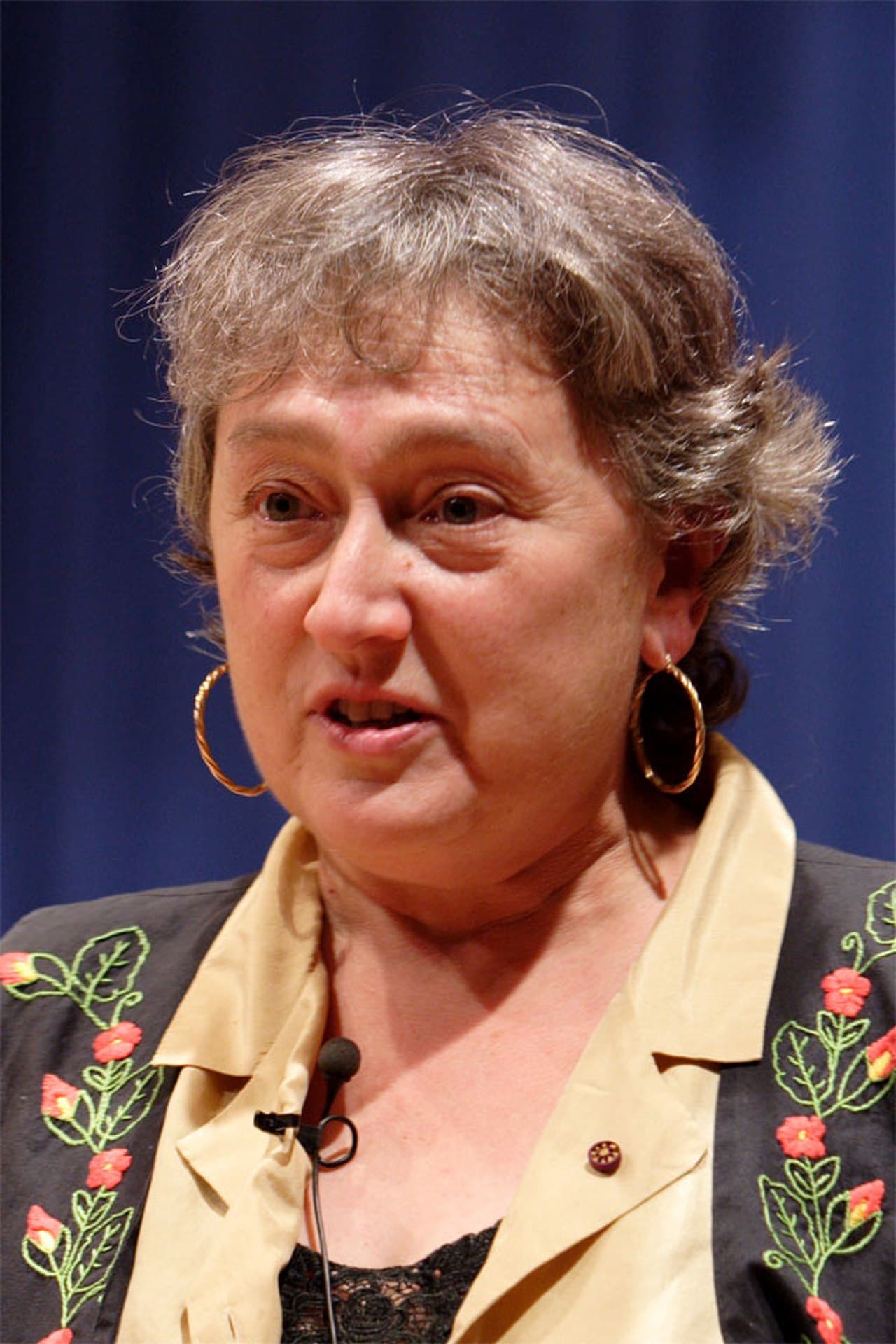
The notion that mitochondria and chloroplasts originated as free-living bacteria was not a sudden epiphany but an idea that germinated over decades. The roots of this hypothesis can be traced back to the observations of German biologist Andreas Schimper in 1883. Schimper noted the striking resemblance between plant chloroplasts and cyanobacteria. Building upon these observations, Russian biologist Konstantin Mereschkowski formalised the concept in 1905, dubbing it 'symbiogenesis'. Despite these early insights, the scientific community largely ignored these propositions. It wasn't until 1967 that a young graduate student, Lynn Margulis, revitalised this idea and integrated it into a comprehensive theory. Her paper, "On the origin of mitosing cells," published in the Journal of Theoretical Biology, faced considerable resistance, enduring rejection from about fifteen journals before finding a home.
Margulis, then Lynn Sagan, was undeterred by the scepticism she encountered. Her hypothesis posited that the organelles responsible for energy production in eukaryotic cells were once independent bacterial entities, engulfed by ancestral host cells. Through her persistence, Margulis presented a radical shift from traditional views of cellular evolution, challenging established norms and laying the groundwork for a profound understanding of the complex interplay between different life forms.
Why it was rejected
The mid-20th-century biological community was deeply entrenched in the principles of the modern synthesis, which emphasised natural selection acting on individual organisms with vertically-inherited genes. In this framework, Lynn Margulis’s proposition appeared to be an extravagant deviation. Her claim required acceptance of horizontal gene transfer at the cellular level, suggesting that one organism could engulf another, and subsequently the two would evolve together as a unified entity. Such a notion was, at the time, viewed as unnecessarily complex and unsupported by existing models of genetic inheritance.
The prevailing explanation suggested that mitochondria were simply specialised compartments that had evolved from the membranes of ancestral eukaryotic cells. To biologists of the era, this explanation was sufficient; it did not necessitate the additional explanatory layers that endosymbiosis introduced. Margulis’s theory required the scientific community to re-evaluate their understanding of evolution and cellular complexity, which was no small ask in a field where the existing paradigm seemed to provide satisfactory answers.
What the evidence eventually showed
It took a series of compelling evidences over the following decades to shift the consensus toward Margulis’s view. The sequencing of mitochondrial DNA in the late 1970s revealed an unmistakable bacterial ancestry, specifically linking mitochondria to the alphaproteobacterial lineage, with modern Rickettsia species among their closest relatives. This genetic evidence was a powerful indicator of their ancient bacterial origins, aligning with Margulis's assertions.
Additionally, the structural and functional characteristics of mitochondrial ribosomes drew further parallels to bacterial counterparts, particularly their susceptibility to antibiotics such as streptomycin, which inhibit bacterial ribosomes but do not affect those of eukaryotic cells. Another point of evidence lay in the mode of reproduction: mitochondria replicate through binary fission, akin to bacterial division, rather than being formed de novo from the eukaryotic cell’s organelles. Chloroplasts, too, exhibit similar patterns, reinforcing the endosymbiotic theory with their clear cyanobacterial lineage. By the 1980s, these converging lines of evidence had turned the tide, establishing endosymbiosis as a cornerstone of modern biology.
When it happened
The mitochondrial endosymbiosis event, estimated to have occurred between 1.5 to 2 billion years ago, followed the Great Oxygenation Event—a pivotal period when atmospheric oxygen levels surged due to photosynthetic activity. This event likely set the stage for an archaeal host cell to engulf, or possibly be invaded by, an alphaproteobacterial cell. What began as a chance occurrence between two cells led to a symbiotic relationship that spanned hundreds of millions of years.
Over this extensive period, the engulfed bacteria transferred the majority of their genes to the host cell’s nucleus, relinquishing their independence and becoming reliant on the host for survival, except for their role in energy production. Chloroplast endosymbiosis, involving a cyanobacterium and an early eukaryotic cell, took place later, approximately 1 to 1.5 billion years ago. Together, these events were transformative, providing the essential energy-generating structures that underpin all complex life forms today.
What still bacterial about mitochondria
Despite their integral role in eukaryotic cells, mitochondria retain several distinctly bacterial traits that echo their evolutionary past. Firstly, they maintain their own genome, albeit reduced, consisting of 37 genes in humans, with 13 encoding essential proteins. Secondly, their protein-synthesis machinery is unique, operating independently from that of the host cell. Their inner membranes possess biochemical features akin to bacterial membranes, such as the presence of cardiolipin, a lipid rare in eukaryotes but common in bacteria.
Mitochondria also divide autonomously from the eukaryotic cell cycle, adjusting their numbers in response to the cell’s energy demands. Furthermore, their mode of inheritance is maternal; paternal mitochondria, delivered via sperm, are typically marked and degraded post-fertilisation, ensuring that mitochondrial DNA is passed exclusively through the maternal line. This pattern of inheritance provides a valuable tool for tracing maternal ancestry, forming the basis for studies on mitochondrial Eve and various population genetics applications.
Margulis's legacy
Lynn Margulis continued to expand the boundaries of biological thought long after her original thesis was validated. She proposed that other cellular structures, such as flagella and centrioles, might have originated from a similar endosymbiotic process involving spirochete bacteria. However, the evidence supporting this 'spirochete hypothesis' has not gained acceptance within the scientific community, highlighting the challenges of extending the endosymbiotic theory beyond its established scope.
Additionally, Margulis co-developed the Gaia hypothesis with James Lovelock, positing that Earth's biosphere behaves as a self-regulating system. While this concept has gained more traction as a metaphorical framework than as a scientifically testable theory, it underscores Margulis’s penchant for bold, integrative thinking. Upon her passing in 2011, Margulis left behind a complex legacy—a scientist who dared to challenge conventions and, in key respects, was vindicated by the evolving scientific narrative.
Cells within our bodies are mosaics of distinct ancestral lineages: nuclear DNA from ancient eukaryotic ancestors, mitochondrial DNA from ancient alphaproteobacteria, and the DNA of countless microbes that inhabit our bodies. This genetic tapestry challenges traditional notions of individuality, suggesting instead a more nuanced view where organisms are composed of myriad interdependent entities. The 21st-century frontier lies in further exploring these connections, as we begin to unravel the intricate web of life that extends beyond our cells.
References
- Sagan, L. (Margulis, L.). (1967). On the origin of mitosing cells. Journal of Theoretical Biology, 14(3), 225–IN6.
- Margulis, L. (1981). Symbiosis in Cell Evolution. W. H. Freeman.
- Gray, M. W., Burger, G., & Lang, B. F. (1999). Mitochondrial evolution. Science, 283(5407), 1476–1481.
- Andersson, S. G. E., et al. (1998). The genome sequence of Rickettsia prowazekii and the origin of mitochondria. Nature, 396, 133–140.